
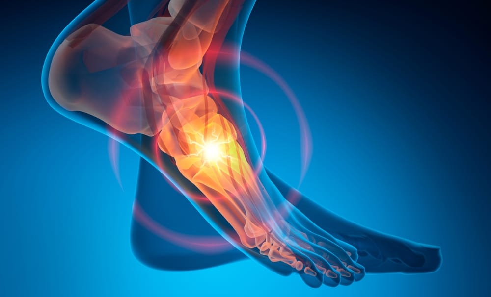
Stress injuries of the foot and ankle are something I see frequently in my clinic,particularly in runners, active individuals, and anyone who has recently increased their training levels. These injuries can be frustrating, often developing gradually and sometimes going undiagnosed for weeks.
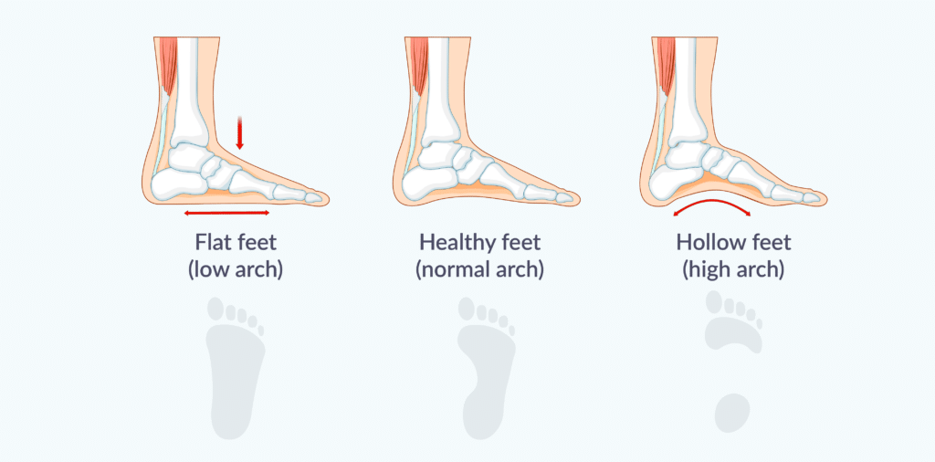
A key factor that is often overlooked is foot biomechanics; in particular, whether you have a flat foot (pes planus) or a high-arched foot (pes cavus).
These structural differences can significantly influence how forces are distributed through the foot and, ultimately, your risk of developing a stress reaction or stress fracture.
In this article, I will explain what stress injuries are, how they develop, the role of foot type, and why the bones themselves are particularly susceptible to these injuries.
Before discussing stress injuries, it is important to understand the bones themselves. The foot and ankle contain 26 bones arranged to provide stability, absorb impact, and allow movement. The ankle joint consists of the tibia, fibula, and talus, while the foot is divided into the hindfoot (talus and calcaneus), midfoot (navicular, cuboid, cuneiforms), and forefoot (metatarsals and phalanges).
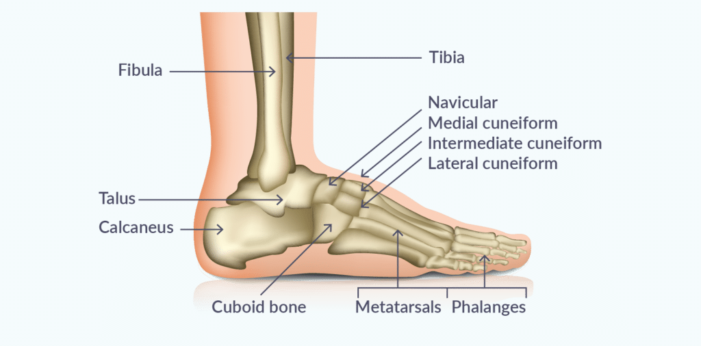
The bones in the foot and ankle are composed of two main layers: the cortical bone, a dense outer shell providing strength, and the trabecular (spongy) bone, which absorbs shock and distributes load. This combination allows the foot to withstand repeated forces, but it also makes certain bones vulnerable.
Areas such as the metatarsals, navicular, talus, and calcaneus are constantly exposed to high mechanical loads during walking, running, and jumping.
If microdamage accumulates faster than the bone can repair itself, this can lead to a stress reaction and, eventually, a stress fracture.
Limited blood supply to some bones, like the navicular or fifth metatarsal, increases the risk of delayed healing or complications if these injuries are not identified early.
Stress injuries exist on a spectrum.
At one end, there is a stress reaction, which is an early-stage injury where the bone becomes inflamed and irritated due to repeated loading.
If this process continues without adequate rest or intervention, it can progress to a stress fracture, where a small crack forms in the bone. Unlike acute fractures caused by a single traumatic event, stress fractures develop gradually due to repetitive mechanical stress exceeding the bone’s ability to repair itself.
Bone is constantly remodelling in response to the forces applied to it. During activity, tiny microdamage occurs naturally and is repaired by the body. Problems arise when:
This imbalance between damage and repair leads to stress injuries.
In my practice, the most commonly affected areas include:
Some bones, like the navicular, are considered high-risk due to their limited blood supply, which can slow healing.
The symptoms often start subtly and worsen gradually.
Patients typically describe:
As the injury progresses, pain may occur earlier during activity and eventually even at rest.
One of the most important contributors to stress fractures is foot shape and biomechanics.
Foot type can influence how forces are distributed across the bones and joints.
A flat foot has a reduced or absent arch, often leading to:
In patients with flat feet, I often see:
Excessive inward motion can place repeated strain on bones that are not designed for prolonged loading in that direction.
High-arched feet tend to be:
In cavus feet, stress is often concentrated rather than dispersed.
As a result, I frequently see:
In simple terms:
Understanding your foot type helps guide both prevention and treatment strategies for stress injuries.
Although foot type and biomechanics are important contributors, stress fractures are usually the result of several risk factors acting together.
This is one of the most common triggers I see in clinical practice.
Increasing running mileage, intensity, frequency, or introducing a new sport too quickly may not give the bone enough time to adapt.
While muscles often strengthen relatively quickly, bone remodelling occurs more slowly, creating an imbalance between the demands being placed on the skeleton and its ability to repair itself.
Footwear plays an important role in how forces are absorbed and transmitted through the foot and ankle.
Shoes that are worn-out, provide inadequate support, or that are inappropriate for your foot type can alter biomechanics and increase loading on specific bones.
In runners and active individuals, simply replacing worn-out shoes or moving to a more suitable style of footwear can sometimes make a significant difference.
Repeated exercise on hard surfaces such as roads, concrete, or artificial pitches exposes the foot and ankle to higher impact forces.
Although the body can adapt to these loads over time, sudden increases in training on harder surfaces can increase the risk of developing a stress reaction or fracture.
Muscles play a critical role in absorbing impact and controlling movement.
When muscles become fatigued or lack sufficient strength, they are less able to dissipate forces effectively.
This means that greater loads are transferred directly to the bones, increasing the risk of stress injury.
Weakness around the foot and ankle, as well as in the hips and core, can all contribute to abnormal loading patterns.
Bone health depends on having sufficient energy and nutrients available for repair and remodelling.
Inadequate calorie intake, restrictive diets, low vitamin D levels, and insufficient calcium intake can all impair bone health and increase susceptibility to stress injuries.
This is particularly important in athletes with high training loads who may inadvertently under-fuel their activity levels.
Bone strength is another important, and sometimes overlooked, factor.
Conditions such as osteopenia (reduced bone density) and osteoporosis (more significant loss of bone strength) make bones less resilient to repetitive loading.
In these situations, even normal training volumes may be sufficient to cause a stress reaction or stress fracture.
This is particularly relevant for women during the perimenopausal and postmenopausal years, when hormonal changes can accelerate bone loss.
The combination of repetitive stress on the skeleton together with reduced bone density can explain why some women become more prone to stress injuries at this stage of life.
In patients with recurrent stress fractures or fractures that seem disproportionate to their activity levels, I will often consider whether an assessment of bone health is appropriate as part of the overall investigation.
In my experience, stress fractures rarely have a single cause.
More commonly, they occur because several factors combine; for example, a runner with a cavus foot who increases training mileage rapidly, wears worn-out shoes, and has underlying osteopenia.
Identifying and addressing all of these contributing factors is essential not only for healing the current injury but also for preventing it from happening again.
Stress fractures can be difficult to spot early, as they may not appear on initial X-rays.
A detailed history and physical examination are essential. I look for:
Early and accurate diagnosis prevents progression and speeds recovery.
Treatment depends on the severity and location of the injury.
Most stress injuries heal without surgery if detected early. Key components of treatment include:
Physiotherapy plays an important role in both recovery and prevention of future stress injuries.
When I see patients with stress reactions or stress fractures, the focus is not only on healing the bone, but also on optimising the mechanics and strength of the foot and ankle to prevent recurrence.
I work closely with a number of highly skilled Physiotherapists, or am very happy to support a physio you are already working with. This multidisciplinary approach ensures that your recovery is as seamless and efficient as possible.
A typical physiotherapy programme will usually focus on:
Initially, patients may need a period of partial or protected weight-bearing, sometimes using crutches, boots, or orthoses.
The aim is to allow the bone to heal while maintaining mobility in surrounding joints.
Once it’s safe to begin loading, we focus on:
Stress injuries often occur when the foot and ankle cannot respond effectively to uneven surfaces. Balance and proprioception training helps restore neuromuscular control, reducing the risk of reinjury.
Physiotherapists may analyse your walking and running mechanics to identify patterns that overload specific bones. Modifying stride, cadence, or foot strike can distribute forces more evenly and protect vulnerable bones.
A structured, stepwise return to running or sport is essential. Jumping or high-impact activities are reintroduced progressively to ensure the bone remodels safely under load.
In my experience, patients who commit to a structured physiotherapy programme not only recover faster but are much less likely to develop recurrent stress injuries.
Because foot shape and biomechanics can play such an important role in the development of stress injuries, I believe podiatry is an essential part of the multidisciplinary team (MDT) approach.
Podiatrists are experts in the assessment of foot function, gait, and lower limb biomechanics. They evaluate how your foot moves during walking and running, identify abnormal loading patterns, and determine whether factors such as flat feet, high arches, limited joint mobility, or muscle imbalances may be contributing to excessive stress on certain bones.
Treatment may involve:
By addressing these underlying mechanical factors, podiatry input not only supports recovery from a stress reaction or stress fracture but also plays a vital role in reducing the risk of recurrence.
Surgery is reserved for:
Surgery aims to restore bone integrity and allow a safe return to activity.
Recovery depends on injury type and severity:
Returning to activity too early is a common cause of recurrence, so gradual progression is essential.
I recommend specialist assessment if you notice:
Early assessment can prevent progression from a stress reaction to a fracture and reduce recovery time.
Stress reactions and stress fractures are not simply overuse injuries, they are the result of a combination of training load, recovery, and biomechanics.
Foot type plays a significant role in determining how forces are distributed through the foot and ankle, influencing the risk and location of injury.
With early and accurate diagnosis, appropriate management, and attention to underlying mechanics, most patients make a full recovery and return to their previous activity levels. If you are struggling with persistent foot or ankle pain, a specialist assessment is the most effective way to identify the cause and guide an effective treatment plan.
Mr Martin Klinke is one of London’s most trusted, and experienced foot specialists. He performs many bunion surgeries each year, and is a highly skilled surgeon.
He offers this surgical treatment to private self-funded and insured patients at the Cleveland Hospital and the Cleveland Clinic in London.
You can find all his patient reviews here.
Fredericson, M., Jennings, F., Beaulieu, C. and Matheson, G.O., 2006. Stress fractures in athletes. Topics in Magnetic Resonance Imaging, 17(5), pp.309-325.
Warden SJ, Burr DB, Brukner PD. Stress fractures: pathophysiology, epidemiology, and risk factors. Current osteoporosis reports,. 2006;4(3):103–109.
Boden, B.P. and Osbahr, D.C., 2000. High-risk stress fractures: evaluation and treatment. JAAOS-Journal of the American Academy of Orthopaedic Surgeons, 8(6), pp.344-353.
Nattiv, A., Kennedy, G., Barrack, M.T., Abdelkerim, A., Goolsby, M.A., Arends, J.C. and Seeger, L.L., 2013. Correlation of MRI grading of bone stress injuries with clinical risk factors and return to play: a 5-year prospective study in collegiate track and field athletes. The American journal of sports medicine, 41(8), pp.1930-1941
Matheson, G.O., Clement, D.B., McKenzie, D.C., Taunton, J.E., Lloyd-Smith, D.R. and MacIntyre, J.G., 1987. Stress fractures in athletes: a study of 320 cases. The American journal of sports medicine, 15(1), pp.46-58.
