
As a foot and ankle surgeon, I often meet patients who’ve been struggling with persistent pain around the back of the foot; sometimes for months, often for years.
The hindfoot (the area including the heel bone and joints just in front of it) plays an important role in walking, running and standing. When something’s not quite right here, it can really affect your mobility, comfort, and activity levels.
So what kind of problems affect the hindfoot, and when might surgery be the right option?
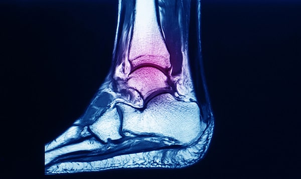
The ankle joint, also known as the talocrural joint, is a hinge joint formed where the tibia and fibula (the two bones of the lower leg) meet the talus bone of the foot. This joint allows the up-and-down movement of the foot (dorsiflexion and plantarflexion), which is essential for walking and running.
Just beneath the ankle lies the hindfoot, made up of the talus and the calcaneus (heel bone). These two bones form the subtalar joint, which enables side-to-side motion (inversion and eversion) and plays a critical role in adapting to uneven surfaces and maintaining balance.
Together, the ankle and hindfoot act as a dynamic unit that absorbs shock, facilitates propulsion, and stabilises the leg during movement. When any part of this complex structure is damaged, be it bone, joint, or surrounding soft tissue, it can have a significant impact on overall mobility and function.

The foot is divided into three sections: the forefoot - toes and metatarsals, midfoot – the arch and its supporting bones, and the hindfoot.
The hindfoot consists primarily of two bones:
These bones form important joints:
This part of the foot is responsible for translating the rotational movement of the leg into foot motion.
It also absorbs ground reaction forces and helps stabilise the body during standing and dynamic movements.
When the hindfoot isn't functioning properly, due to injury, arthritis, or deformity - it often leads to compensatory issues higher up the chain, including knee, hip, or back pain.
Patients with hindfoot pathology tend to describe a fairly typical set of symptoms, although the exact pattern can vary depending on the underlying condition.
Some of the things I frequently hear in clinic include:
One gentleman I saw recently described a ‘persistent pain on the inside of the ankle and foot’ and a ‘rolling-in’ sensation whenever he walked, and he had also started to notice his trainers wearing unevenly. This ultimately turned out to be early-stage posterior tibial tendon dysfunction.
A big part of my job as a foot and ankle specialist, is being able to make an accurate diagnosis, in order to determine the best cause of action; whether surgery is appropriate, or whether the problem can be managed conservatively.
All my assessments starts with a thorough history and physical examination.
In clinic, I will typically:
Within the facilities that I work, I have excellent access to imaging test, which can help to confirm the diagnosis:
Only after a full clinical picture is built can we confidently determine what the diagnosis and prognosis is, and whether surgery is likely to be needed.
This decision process is always patient led, and discussed in full. Every patient and case is different; I help to facilitate that informed, decision making process.
While many hindfoot issues can be managed without surgery, especially if caught early, however some conditions progress to the point where an operation becomes the most effective option.
Here are some of the most frequent causes of hindfoot pain that I see in clinic, and how we approach treatment:

PTTD is one of the most common causes of adult-acquired flatfoot. It occurs when the posterior tibial tendon - which runs along the inside of the ankle and supports the arch - becomes inflamed, overstretched or torn.
This can result from overuse, trauma, or age-related degeneration. Left untreated, the arch collapses, the heel begins to drift outward, and the foot becomes increasingly rigid and painful.
In the early stages (known as Stage I), non-surgical treatments can be very effective:
However, once the injury develops and the foot becomes fixed in a deformed position (Stage II, III or IV), surgery is usually required.
Procedures may include:
Arthritis may involve one or more joints, most commonly the subtalar joint (between the talus and heel bone), the talonavicular joint, the calcaneocuboid joint, or the ankle joint itself (talocrural joint).
Arthritis can result from osteoarthritis (wear and tear), rheumatoid arthritis (inflammatory), or post-traumatic arthritis following previous injuries such as fractures or ligament damage.
Symptoms typically include:
Early treatment focuses on:
If symptoms persist despite these measures, surgical options include:
If conservative treatment fails and quality of life is significantly affected, surgery may be considered. The choice of surgery depends on which joints are involved, the extent of damage, and the patient’s activity level.
Surgical options include:
Tarsal coalition is a congenital condition in which two or more bones in the hindfoot are abnormally connected, usually by fibrous, cartilaginous or bony tissue. It’s often diagnosed in adolescence, when the previously flexible foot starts to stiffen, causing pain and recurrent ankle sprains.
Symptoms may include:
Initial management includes:
Unfortunately, not everyone responds to conservative treatment, so when symptoms persist or the coalition significantly restricts movement and quality of life, surgery may be recommended.
Tarsal Coalition Surgery
Depending on the type and location of the coalition, options include:
With the right procedure and rehab, many patients regain good mobility and return to sports or daily activity without pain.

Structural deformities such as flatfoot (pes planus) or high arch foot (pes cavus) can cause abnormal loading through the hindfoot, leading to pain, imbalance, and degeneration of joints over time.
Flatfoot deformity is often progressive and may be caused by PTTD, arthritis, or congenital issues. If left unaddressed, it can lead to hindfoot valgus (outward heel tilt), midfoot collapse, and forefoot abduction.
Conversely, cavus foot causes the heel to tilt inward (hindfoot varus), leading to pressure on the outer foot and frequent ankle sprains.
Symptoms include:
Conservative treatments may include:
When non-surgical measures are no longer effective, surgery aims to realign the hindfoot, correct soft tissue imbalance, and offload painful joints.
Surgical options include:
Each surgical plan is tailored to the individual – there is no one size fits all, and I make sure that surgery is only used as a last resort and when it is in the best interest of my patients.
Hindfoot surgery is not a quick fix. Most procedures require a period of non-weightbearing, followed by gradual rehabilitation. I always make this very clear to patients. But if you’re prepared for the recovery journey, the improvement in pain and function can be transformative and life-changing.
Every foot is different. And every patient has different goals; whether it’s walking the dog again without limping, chasing after grandchildren, getting back to five-a-side football, or back to elite level sport.
If you’ve been living with ongoing hindfoot pain or deformity, it’s worth having a proper assessment. Often, the right diagnosis and early intervention - whether surgical or not - can spare you months or years of discomfort.
If you have any concerns or questions, I’d be happy to see you in clinic.
Mr Martin Klinke is one of London’s most trusted, and experienced foot specialists. He performs many bunion surgeries each year, and is a highly skilled surgeon.
He offers this surgical treatment to private self-funded and insured patients at the Cleveland Hospital and the Cleveland Clinic in London.
You can find all his patient reviews here.