Managing Hindfoot Pain and Arthritis During Cold Weather

Cold, damp weather has a funny way of revealing old injuries. Every winter I see a rise in patients coming in with painful, stiff feet and ankles – particularly those with arthritis in the hindfoot.

It’s that deep, dull ache that’s worse in the mornings, eases a bit as you move, then flares up again after a walk in the cold. Sound familiar?

Let’s talk about why this happens, what’s going on inside the foot, and most importantly, what you can do about it; from simple adjustments to footwear to surgical options that can make a world of difference.

Understanding the Hindfoot

The hindfoot is the back section of the foot – the bit that connects your ankle to the midfoot. It includes the talus (the bone that forms the lower part of the ankle joint) and the calcaneus, or heel bone. Between them sits the subtalar joint, which allows your foot to roll inwards and outwards – essential for walking on uneven ground and for shock absorption.

When arthritis affects the subtalar or talonavicular joints, the smooth cartilage that cushions movement becomes worn or damaged. Without that cartilage, bone rubs against bone, leading to stiffness, swelling, and pain.

Why Cold Weather Makes It Worse

Many patients tell me their foot feels fine in summer but “locks up” in winter. There’s actually some logic behind this.

Cold and damp conditions can cause the tissues around joints, particularly synovial fluid and tendons, to become less elastic. That means your joints feel stiffer, less lubricated, and slower to get going.

Research backs this up: around 70% of people with osteoarthritis report weather-related changes in symptoms, especially when temperatures drop or barometric pressure changes.

Although we can’t control the climate, understanding the triggers helps you plan ahead – and manage your condition more effectively.

Who Gets Hindfoot Arthritis?

Hindfoot arthritis isn’t just a problem for older adults. I see it in a wide range of patients, from retired walkers to active runners and even footballers.

The common causes include:

In my clinic, I’d say around 1 in 5 patients I see with chronic foot pain have some degree of hindfoot or subtalar joint arthritis – and the number seems to rise each year as people stay more active later in life.

Common Symptoms of Hindfoot Arthritis

Patients usually describe:

I often see people who say they’ve been “putting up with it” for years, thinking it’s just part of ageing – until the winter months make it unbearable.

Assessing the Problem

Diagnosis starts with a good clinical assessment. I’ll often test how much movement there is through the subtalar joint – it should glide smoothly from side to side. Loss of that movement, with tenderness and swelling, is usually a telltale sign.

Imaging helps confirm it:

Managing Hindfoot Arthritis Conservatively

When symptoms are mild to moderate, conservative treatment can be highly effective. The goal is to reduce pain, improve mobility, and slow progression of joint damage.

Footwear and Orthotics

Shoes make a huge difference. I often recommend stiff-soled walking shoes or trainers with a rocker-bottom sole, which reduce the need for painful side-to-side motion at the subtalar joint.

Custom orthotics can correct alignment and offload pressure points – particularly useful if your pain stems from flat feet or previous injury. I recently had a patient, a 62-year-old keen rambler, who could barely manage a 2-mile walk before pain kicked in. With the right orthotic inserts and supportive boots, she’s now comfortably hiking 6–8 miles again.

Activity Modification

Switching to low-impact activities such as cycling, swimming, or using an elliptical trainer can maintain fitness without aggravating the joint. Even walking poles can reduce strain on uneven ground.

Physiotherapy

Physio focuses on strengthening surrounding muscles and improving balance. Gentle range-of-motion exercises help maintain mobility without overloading the joint.

Injections and Medications

Corticosteroid injections
A corticosteroid injection is essentially a strong, targeted anti-inflammatory medicine delivered directly into the painful joint.

By targeting the inflammation at its source, they can provide excellent pain relief, often for several months, and in some cases years.

They are especially effective during acute flare-ups, when inflammation within the joint is driving most of the pain. We will confirm this using MRI imaging prior to progressing with treatment.

Some patients only need one injection; others might benefit from occasional repeat injections during bad flare-ups.

Platelet-rich plasma (PRP) injections

For those looking for a more biological or ‘regenerative’ option, PRP injections are becoming increasingly popular. PRP is made from your own blood; a small sample is taken, spun in a centrifuge to concentrate the platelets, and inject the plasma back into the joint. Platelets are rich in growth factors, which may help calm inflammation and support tissue healing.

The evidence for PRP is still evolving, particularly for hindfoot and subtalar arthritis. Some patients report meaningful improvement, while others notice only mild or temporary benefit.

Anti-inflammatory medication
Anti-inflammatory tablets (NSAIDs) can also help settle symptoms during a flare-up. They aren’t a long-term fix, but they do a good job at reducing pain and swelling in the short term. They’re not suitable for everyone, particularly people with stomach, kidney or heart problems, so I always advise using them under guidance and for limited periods only.

Lifestyle and Holistic Management

Cold weather care matters. Keeping your feet warm, using thermal socks, and doing gentle mobility exercises before heading out can make a big difference.

Maintaining a healthy weight also reduces load through the hindfoot – if you have a high BMI, losing even 10% of your bodyweight can noticeably ease symptoms.

When Conservative Measures Aren’t Enough

For some patients, no matter how supportive the footwear or how diligent the physio, the pain still limits everyday life. When arthritis becomes end-stage – where cartilage is completely worn away – surgery often becomes the most effective route forward.

Surgical Options

During surgery, the damaged cartilage is removed, the bones are aligned, and small screws hold them together while they fuse.

Outcomes and Recovery

Fusion surgery has a success rate of around 90 - 95% for pain relief and improved function. The trade-off is a small loss of side-to-side motion, but most patients find this barely noticeable compared to the improvement in comfort.

Recovery requires patience. You’ll typically be in a cast or boot for 6 - 8 weeks, keeping weight off the foot initially to allow the bones to fuse. Gradual weight-bearing starts once healing is confirmed on X-ray, followed by physiotherapy to restore strength and balance.

Most people return to comfortable walking by around 3 - 4 months, and to more active pursuits like hiking or golf by 6 - 9 months. One of my patients summed it up nicely:

“I’d forgotten what it felt like to walk without wincing. The surgery gave me my life back.”

That’s the sort of result that makes the decision worthwhile.

Final Words

Living with hindfoot arthritis, especially through the cold, damp British winter – can be frustrating. The stiffness, the ache that creeps up when the temperature drops, the way uneven ground suddenly feels like a mountain. But the good news is there’s a lot we can do.

From the right footwear and orthotics to injections, physiotherapy, and surgical solutions, there’s a full spectrum of treatment options available to you.

The key is recognising when conservative measures have done all they can, and being open to surgery if that’s what’s needed to restore quality of life.

So, if your hindfoot pain is holding you back this winter, don’t just grin and bear it.

Book an assessment with me, and let’s get you moving comfortably again.

Q&As

What exactly is subtalar (hindfoot) arthritis?
Subtalar arthritis is wear-and-tear (or inflammatory) damage to the cartilage and surfaces of the subtalar joint - the joint between the talus and calcaneus in the hindfoot. It causes deep pain in the heel/behind the ankle, stiffness when walking on uneven ground, and sometimes swelling.

Why does my hindfoot hurt more in cold and damp weather?
Many patients report weather-related worsening. Systematic reviews show a statistically significant association between weather factors (temperature, humidity, barometric pressure) and OA pain in many studies, although findings are not uniform across every paper. It’s reasonable to acknowledge the symptom pattern and advise practical measures (warmth, footwear, gentle mobilisation).

Can orthotics or special shoes really help?
Yes, supportive footwear, rocker-soled shoes and targeted orthoses can reduce painful subtalar motion and redistribute load across the foot. Clinical and biomechanical studies demonstrate reduced painful motion and symptomatic benefit in selected patients. Orthotics are a low-risk first line and often worth a trial.

Are injections (steroid or PRP) useful for hindfoot arthritis?
Corticosteroid injections often give meaningful short-term relief by reducing inflammation inside the joint. PRP has been investigated: meta-analyses and RCT data are mixed - some short-term improvements have been reported, but a high-quality RCT did not show significant benefit at 26 weeks for ankle OA. I will always discuss realistic expectations and evidence with you before we make a decision on treatment.

If I have surgery (subtalar fusion), what are realistic outcomes and recovery times?
Subtalar fusion is the standard for end-stage subtalar arthritis and has high rates of pain relief (many series report >85–90% good outcomes).

Expect a period of protected weight-bearing in a cast/boot for about 6 - 8 weeks, progressive physiotherapy, comfortable walking by 3 - 4 months and return to most activities (including light hiking/golf) by c. 6 - 9 months depending on individual healing.

Suggested reading

  1. Joveniaux, P., Philippot, R., Ardilouze, P., Hardy, P., & Sautet, A. (2010) ‘Long-term results of in situ subtalar arthrodesis’, Foot & Ankle International, 31(4), pp. 329–335.
  2. Wang, L., Xie, H., Li, Z., et al. (2023) ‘Association between weather factors and osteoarthritis pain: a systematic review and meta-analysis’, Osteoarthritis and Cartilage (systematic review), 31(7), pp. 1000–1013.
  3. Riskowski, J.L., Hagedorn, T.J., Dufour, A.B., et al. (2011) ‘Arthritis, foot pain and shoe wear: current musculoskeletal evidence’, Journal of Foot & Ankle Research, 4:11.
  4. Huang, Y.C., et al. (2006) ‘Effects of ankle-foot orthoses on ankle and subtalar biomechanics during gait’, Gait & Posture, 24(3), pp. 272–281.
  5. Paget, L.D.A., et al. (2019) ‘Platelet-Rich plasma Injection Management for Ankle OA (PRIMA): protocol for a randomised, double-blind, placebo-controlled trial’, BMJ Open, 9:e030961.
  6. Paget, L.D.A., et al. (2021) ‘Effect of platelet-rich plasma injections versus placebo on ankle osteoarthritis symptoms: a randomized clinical trial’, JAMA (RCT), 326(10), pp. 949–957. (PRP ankle RCT showing no significant benefit at 26 weeks).
  7. British Orthopaedic Association (BOA) (2016) ‘Hindfoot arthritis: guidance on management’ (clinical summary/guideline).

Understanding the Difference Between a Bunion and Big Toe Arthritis

Pain around the big toe joint is one of the most common reasons patients come to see me in clinic. For some, the problem is a gradually enlarging bump at the base of the toe that rubs against shoes. For others, it’s stiffness, aching, and swelling that seem to worsen with every step.

Although these symptoms often overlap, the causes can be quite different - the two most frequent culprits being bunions and big toe arthritis.

Understanding which you’re dealing with is key to getting the right treatment and keeping you comfortably on your feet.

What is a bunion?

A bunion (hallux valgus) is a structural deformity of the big toe joint, where the toe begins to drift towards its neighbour and a bony bump develops on the inside of the foot. Over time, this misalignment can cause pressure, irritation, and discomfort — particularly in tight or narrow shoes.

In my practice, I often see bunions that have been slowly progressing for years. Many patients tell me they first noticed a slight bump that only became painful later on. Bunions can run in families and are often linked to inherited foot shape, but poorly fitting footwear can certainly accelerate their development.

What is big toe arthritis?

Big toe arthritis is a different condition altogether. Rather than a visible bump on the side of the foot, the problem lies within the joint itself. The smooth cartilage that allows the big toe to move freely becomes worn down, leading to pain, stiffness, and swelling. Some patients describe a “grinding” or “catching” sensation when they try to bend their toe.

Unlike bunions, big toe arthritis often causes difficulty during push-off when walking or running, and the pain tends to worsen with activity rather than shoe pressure alone.

How can you tell them apart?

A bunion typically presents as a visible bump at the base of the big toe, with the toe angling inwards.

In contrast, arthritis often causes a stiff, swollen joint with little outward deformity. Patients with arthritis tend to notice a gradual loss of motion (Hallux Rigidus) and discomfort even in roomy footwear.

In hallux rigidus there sometimes is a bump, but it is usually more on the top of the joint due to bony growth. It’s something patients can feel, which may cause trouble in shoes.

During a consultation, I’ll assess alignment, mobility, and tenderness, often supported by an X-ray. This helps confirm whether we’re dealing with a structural deformity or joint wear and tear - and guides how best to manage it.

Treatment: finding the right balance

For both bunions and arthritis, we always start with non-surgical management.

That said, when pain persists or starts to limit daily activities, it’s time to consider surgical options.

Surgical options for bunions

When conservative treatments no longer relieve symptoms, bunion surgery can be highly effective. The aim is not just to remove the bump but to realign the bones and restore the natural shape of the foot.

There are different surgical ways of correcting a bunion and the type of operation will be adjusted depending on the severity of the bunion.

The most common approach I perform is an osteotomy, where the bone is precisely cut and repositioned before being fixed with small screws. This restores alignment and allows the joint to function more normally.

In more advanced cases, particularly where arthritis has also developed, a fusion (arthrodesis) of the joint may be the best way to eliminate pain and stabilise the toe.

Recovery typically involves a period of protected weight-bearing in a special shoe, followed by gradual return to normal footwear. Most of my patients are walking comfortably again within a few weeks - and often remark how much better their foot feels once the pressure and deformity are gone.

Surgical options for big toe arthritis

For big toe arthritis, the choice of surgery depends on how much cartilage has been lost. In earlier stages, joint-preserving procedures can be performed; this involves removing small bone spurs and freeing up movement in the joint. It’s a relatively simple operation with quick recovery and excellent results in suitable cases.

For more advanced arthritis, where the joint surfaces are severely worn, I often recommend a joint fusion (arthrodesis). This procedure permanently joins the bones of the big toe joint together, removing pain completely and restoring stability.

Although you lose some movement, most patients find they can walk, hike, and even run without discomfort after recovery.

In certain cases, particularly for lower-impact lifestyles, a joint replacement (arthroplasty) may be an option to preserve motion.

A final word

Both bunions and big toe arthritis are common - and I see them in everyone from active runners to patients who simply want to enjoy walking without pain. The key is early assessment and tailored treatment.

If you’re struggling with pain, stiffness, or deformity around your big toe, don’t ignore it. A careful clinical assessment and imaging can quickly clarify the cause, and with the right approach, whether it’s simple footwear advice or a modern surgical correction, there’s every reason to expect a smooth, active recovery.

Patient Q&A Section

Q1: How do I know if I have a bunion or big toe arthritis?
A bunion causes a visible bump on the side of the big toe and is often irritated by tight shoes. Big toe arthritis, however, causes stiffness and deep joint pain even in roomy footwear. An X-ray and clinical examination are the best ways to confirm which condition you have.

Q2: Do bunions always need surgery?
Not always. Many patients manage their bunion pain successfully with wide shoes, orthotics, and activity modification. Surgery is considered if pain persists or the deformity worsens despite conservative care.

Q3: What are the signs that I might need surgery for big toe arthritis?
If your toe becomes increasingly stiff or painful, especially during walking or exercise, and non-surgical treatments are no longer effective, it may be time to discuss surgical options such as cheilectomy, joint fusion, or replacement.

Q4: How long is recovery after bunion surgery?
Most patients can walk in a protective shoe immediately after surgery and transition to normal footwear within 6 - 8 weeks. Full recovery, including swelling reduction and return to sport, can take a few months depending on the procedure performed.

Q5: Can bunions and arthritis occur together?
Yes. Long-standing bunions can sometimes lead to arthritic changes in the big toe joint. This is why early assessment and management are important - to prevent progressive joint damage.

NHS – Bunions Overview:
https://www.nhs.uk/conditions/bunions/
(Symptoms, causes, treatment, and when to see a specialist.)

NHS – Osteoarthritis:
https://www.nhs.uk/conditions/osteoarthritis/
(Information on arthritis management and self-care.)

Shockwave Therapy for Heel and Ankle Pain - A Non-Invasive Solution

When patients come to see me with heel or ankle pain, one of the first things they usually ask is: “Do I need surgery?”

The good news is that in most cases, the answer is no. Over the years, we’ve made huge progress in managing painful conditions such as plantar fasciitis, Achilles tendinopathy, and certain ankle problems without resorting to invasive procedures.

One of the most effective tools we now have in our toolkit is shockwave therapy.

With marathon and half-marathon season in full swing this autumn, I’m seeing an increasing number of runners and active people struggling with stubborn pain that just won’t shift.

But it’s not just athletes; many of my patients are people who are on their feet all day, or those who simply enjoy walking but find themselves sidelined by pain. That’s where shockwave therapy can make all the difference.

What Is Shockwave Therapy?

Despite its name, shockwave therapy has nothing to do with electricity. Instead, it involves delivering high-energy sound waves directly to the injured tissue. These waves stimulate the body’s natural healing response, increasing blood flow and encouraging the repair of damaged tissues.

The treatment is carried out in clinic, with no need for anaesthetic or hospital admission. It usually takes only 10–15 minutes per session, and patients can walk out straight afterwards. Most people require a course of three to five sessions, spaced a week apart, to see the best results.

Conditions We Commonly Treat

Shockwave therapy has been particularly successful in treating stubborn, long-standing conditions that haven’t improved with rest, physiotherapy, or standard non-surgical treatments. The main ones I see in clinic are:

These conditions can be incredibly frustrating, particularly for runners preparing for a race or for people who simply want to stay active without pain. Shockwave therapy gives us a way of tackling the problem head-on without invasive surgery.

Why Shockwave Therapy?

There are several reasons why I recommend shockwave therapy for the right patients:

What Does It Feel Like?

A common concern patients raise is whether the treatment hurts. The sensation can be uncomfortable; like a series of sharp taps on the skin—but it is usually very tolerable. The intensity can be adjusted to ensure it’s manageable, and most patients describe it as “odd” rather than painful.

Importantly, because the treatment is stimulating your body’s healing response, the benefits often build gradually over the weeks following treatment rather than being instant. Patience really does pay off here.

Shockwave Therapy and the Marathon Season

Each year as autumn approaches, I see a wave of patients training for marathons and half-marathons. Many are nursing small injuries or overuse problems, particularly in the feet and ankles.

Training plans often involve steadily increasing mileage, which places repetitive stress on the Achilles tendon and plantar fascia. If these tissues aren’t given enough time to adapt, micro-injuries accumulate and pain develops.

For some runners, this pain lingers long after the race, or even forces them to stop training altogether.

Shockwave therapy is especially valuable in these cases. It can help athletes manage their pain, recover more effectively, and return to training sooner. Combined with a structured rehabilitation plan and load management advice, it offers a way back to running that doesn’t involve months off or surgery.

Who Is Shockwave Therapy Suitable For?

While shockwave therapy is not a magic bullet, it can be life-changing for the right patients. It tends to work best for those who:

It is not usually the first-line treatment; I often recommend physiotherapy, footwear changes, and activity modification first. But if those haven’t worked, shockwave therapy is an excellent next step.

Patient FAQs

How many sessions will I need?
Most patients require between four and six sessions, usually one per week.

Will I need time off work or sport?
No, most people continue with daily activities. You may need to reduce high-impact training temporarily, but complete rest is rarely required.

Is it safe?
Yes. Shockwave therapy is safe when performed by a trained specialist. Side effects are usually mild, such as short-lived redness, swelling, or bruising.

Does it work straight away?
Not always. Many patients notice gradual improvement over several treatments, as the body’s healing response builds.

Can it prevent me needing surgery?
In many cases, yes. For persistent heel or Achilles pain, shockwave therapy often provides enough relief to avoid surgery altogether.

My Final Thoughts

Heel and ankle pain can be incredibly disruptive; whether you’re preparing for a marathon, trying to keep up with grandchildren, or simply walking the dog. Radial shockwave therapy offers a safe, non-invasive, and effective solution for stubborn plantar fasciitis, Achilles tendinopathy, and other ankle pathologies.

As a foot and ankle specialist, I’ve seen many patients who felt they had run out of options finally find relief with this treatment.

If you’ve been struggling with persistent heel or ankle pain, it might be time to consider whether shockwave therapy could help you. Please get in touch to book an appointment.

References

Dimitrios, K. et al. (2016). Radial extracorporeal shockwave therapy for the treatment of chronic plantar fasciopathy: a systematic review and meta-analysis. British Journal of Sports Medicine, 50(16), 972–981.

Gerdesmeyer, L. et al. (2008). Extracorporeal shock wave therapy for the treatment of chronic plantar fasciitis: a randomized controlled multicentre trial. Journal of Bone and Joint Surgery, 90(3), 270–278.

Rompe, J. D. et al. (2009). Shock wave therapy for chronic Achilles tendinopathy: a double-blind, randomized controlled trial. Clinical Orthopaedics and Related Research, 467(12), 3235–3242.

Speed, C. (2014). A systematic review of shockwave therapies in soft tissue conditions: focusing on the evidence. British Journal of Sports Medicine, 48(21), 1538–1542.

Morton’s Neuroma - Finding Relief from Nerve Pain in Your Feet

Ever feel like there’s a stone in your shoe, but when you check, there’s nothing there? That sharp, burning sensation or the feeling of numbness between your toes might be a sign of Morton's neuroma.

This often-overlooked nerve condition affects thousands of people, especially those with active lifestyles. But what exactly causes it, and why might you be experiencing it now?

In this blog, we look more closely into the symptoms, causes, and most importantly, how to find relief.

What is Morton’s Neuroma?

Morton’s Neuroma, also known as intermetatarsal neuroma, is a painful condition that affects the ball of the foot, most commonly between the third and fourth toes.

It was first documented by an American surgeon called Thomas George Morton in 1876!It is not actually a tumour but rather a thickening of the tissue surrounding the interdigital nerves, which can lead to nerve irritation and compression.

This condition results in sharp, burning pain, numbness, and tingling in the foot and toes which can be quite severe and make walking or running very painful.

Who Gets Morton’s Neuroma and Why?

Morton’s Neuroma is a relatively common foot condition which tends to affect adults, especially women between the ages of 30 and 60. It rarely affects both feet, certainly not at the same time.

Several factors can increase the likelihood of developing this condition:

Footwear

Wearing tight, high-heeled, or narrow shoes that squeeze the toes can significantly increase pressure on the forefoot. This compression may contribute to nerve irritation over time.

Foot Structure

People with certain foot types, such as flat feet, high arches, or bunions, are more prone to developing Morton’s Neuroma. These structural abnormalities can alter the weight distribution on the feet, placing extra stress on the metatarsal area.

Repetitive Trauma

Individuals who engage in high-impact activities, such as dancing, running, or sports that involve frequent pressure on the forefoot like tennis or basketball, are at a higher risk of developing this condition due to repetitive microtrauma to the nerve.

Pre-existing Conditions

Conditions such as hammertoes, where the toes bend abnormally, can cause additional pressure on the nerves, contributing to the development of Morton’s Neuroma.

Symptoms of Morton’s Neuroma

The primary symptom of Morton’s Neuroma is pain in the ball of the foot. This pain is often described as sharp, burning, or akin to walking on a pebble. Other key symptoms include:

Symptoms tend to fluctuate, often worsening during periods of activity and improving when resting or wearing wider, more comfortable footwear.

Diagnosis of Morton’s Neuroma

Diagnosing Morton’s Neuroma typically begins with a thorough physical examination by a healthcare professional, who will assess the patient's medical history and symptoms. During the examination, the clinician may:

To confirm the diagnosis and rule out other possible conditions such as stress fractures or arthritis, imaging studies may be used:

What is the Best Treatment for Morton’s Neuroma?

Treatment for Morton’s Neuroma depends on the severity of the condition and how long symptoms have been present. The goal is to relieve nerve irritation and reduce pressure on the affected area. Treatment options range from conservative approaches to more invasive interventions:

Conservative Treatment for Morton’s Neuroma

2.      Medications and Injections for Morton’s Neuroma

Will Morton’s Neuroma Ever Go Away?

Morton’s Neuroma does not typically resolve on its own without some form of intervention. However, conservative treatments, such as footwear changes, orthotics, and physiotherapy can help manage symptoms and prevent the condition from worsening.

With appropriate care, many patients find significant relief, and their symptoms may subside. Early diagnosis and treatment increase the chances of a favourable outcome.

For patients who experience chronic symptoms, minimally invasive treatments like injections or surgery may be necessary to provide lasting relief.

Surgical Treatment for Morton’s Neuroma

Surgery for Morton’s Neuroma is a very safe and successful procedure, and is often the best long term solution.

Evidence shows that surgical intervention can offer significant and lasting relief, particularly for patients who experience debilitating pain that impacts daily function. Studies suggest that around 80-90% of patients who undergo a neurectomy report substantial or complete symptom relief post-surgery.

The procedure effectively removes the portion of the nerve causing discomfort, reducing the painful sensations between the toes. Research also indicates that surgical outcomes are long lasting, with most patients experiencing sustained relief even years after the procedure.

Recovery times vary, but many patients return to their normal activities within weeks to a few months, depending on the complexity of the case and adherence to post-operative care. For those who have exhausted non-surgical options, surgery remains a highly effective treatment for restoring foot comfort and functionality.

The two common surgical options are:

What Happens If You Don’t Treat Morton’s Neuroma?

If Morton’s Neuroma is left untreated, the pain and discomfort are likely to worsen over time. This can lead to several complications:

Recent Research on Morton’s Neuroma

Recent studies have focused on understanding the most effective treatment approaches for Morton’s Neuroma. One notable area of research is the use of alcohol sclerosing injections. A 2022 study demonstrated that patients receiving a series of alcohol injections had significant improvements in pain and functional outcomes compared to conservative treatments alone.

Additionally, a study published in 2023 explored the role of shockwave therapy for Morton’s Neuroma, suggesting that this non-invasive approach could reduce pain and inflammation by promoting tissue healing and nerve regeneration.

Conclusion

Morton’s Neuroma is a painful condition that can significantly impact daily life if left untreated. Fortunately, a variety of treatment options exist, ranging from conservative approaches to more invasive procedures. Early diagnosis and intervention are key to managing symptoms and preventing long-term complications. Whether through footwear changes, orthotics, injections, or surgery, patients have a high likelihood of achieving relief and returning to the activities they love.

References

  1. Masala, S., Fiori, R., Raguso, M., et al. (2022). "Percutaneous alcohol injection in the management of Morton’s neuroma: A review of outcomes." Journal of Foot & Ankle Research 15(4), 321–326.
  2. Zhu, M., Yang, L., Li, J., et al. (2023). "Efficacy of alcohol sclerosing injections versus corticosteroid injections for Morton’s neuroma: A randomized trial." International Journal of Foot & Ankle Surgery, 9(1), 11–17.
  3. Ahmed, M., et al. (2022). "Sclerosing injections for treating Morton’s neuroma: A systematic review and meta-analysis." European Journal of Orthopaedic Surgery & Traumatology, 32(5), 819–825.
  4. Koo, Y., Lee, C. H. (2023). "The effects of extracorporeal shockwave therapy in patients with Morton’s Neuroma: A pilot study." Journal of Rehabilitation Research and Development, 60(2), 15-20.

Hindfoot Conditions - When is Surgery Needed? 

As a foot and ankle surgeon, I often meet patients who’ve been struggling with persistent pain around the back of the foot; sometimes for months, often for years.

The hindfoot (the area including the heel bone and joints just in front of it) plays an important role in walking, running and standing. When something’s not quite right here, it can really affect your mobility, comfort, and activity levels.

So what kind of problems affect the hindfoot, and when might surgery be the right option?

Ankle Joint Anatomy

The ankle joint, also known as the talocrural joint, is a hinge joint formed where the tibia and fibula (the two bones of the lower leg) meet the talus bone of the foot. This joint allows the up-and-down movement of the foot (dorsiflexion and plantarflexion), which is essential for walking and running.

Just beneath the ankle lies the hindfoot, made up of the talus and the calcaneus (heel bone). These two bones form the subtalar joint, which enables side-to-side motion (inversion and eversion) and plays a critical role in adapting to uneven surfaces and maintaining balance.

Together, the ankle and hindfoot act as a dynamic unit that absorbs shock, facilitates propulsion, and stabilises the leg during movement. When any part of this complex structure is damaged, be it bone, joint, or surrounding soft tissue, it can have a significant impact on overall mobility and function.

A Closer Look at the Hindfoot

The foot is divided into three sections: the forefoot - toes and metatarsals, midfoot – the arch and its supporting bones, and the hindfoot.

The hindfoot consists primarily of two bones:

These bones form important joints:

This part of the foot is responsible for translating the rotational movement of the leg into foot motion.

It also absorbs ground reaction forces and helps stabilise the body during standing and dynamic movements.

When the hindfoot isn't functioning properly, due to injury, arthritis, or deformity - it often leads to compensatory issues higher up the chain, including knee, hip, or back pain.

Symptoms People Commonly Report

Patients with hindfoot pathology tend to describe a fairly typical set of symptoms, although the exact pattern can vary depending on the underlying condition.

Some of the things I frequently hear in clinic include:

One gentleman I saw recently described a ‘persistent pain on the inside of the ankle and foot’ and a ‘rolling-in’ sensation whenever he walked, and he had also started to notice his trainers wearing unevenly. This ultimately turned out to be early-stage posterior tibial tendon dysfunction.

How is the Hindfoot Assessed?

A big part of my job as a foot and ankle specialist, is being able to make an accurate diagnosis, in order to determine the best cause of action; whether surgery is appropriate, or whether the problem can be managed conservatively.

All my assessments starts with a thorough history and physical examination.

In clinic, I will typically:

Within the facilities that I work, I have excellent access to imaging test, which can help to confirm the diagnosis:

Only after a full clinical picture is built can we confidently determine what the diagnosis and prognosis is, and whether surgery is likely to be needed.

This decision process is always patient led, and discussed in full. Every patient and case is different; I help to facilitate that informed, decision making process.

Common Conditions That May Require Hindfoot Surgery

While many hindfoot issues can be managed without surgery, especially if caught early, however some conditions progress to the point where an operation becomes the most effective option.

Here are some of the most frequent causes of hindfoot pain that I see in clinic, and how we approach treatment:

Posterior Tibial Tendon Dysfunction (PTTD)

PTTD is one of the most common causes of adult-acquired flatfoot. It occurs when the posterior tibial tendon - which runs along the inside of the ankle and supports the arch - becomes inflamed, overstretched or torn.

This can result from overuse, trauma, or age-related degeneration. Left untreated, the arch collapses, the heel begins to drift outward, and the foot becomes increasingly rigid and painful.

In the early stages (known as Stage I), non-surgical treatments can be very effective:

However, once the injury develops and the foot becomes fixed in a deformed position (Stage II, III or IV), surgery is usually required.

Procedures may include:

Hindfoot Arthritis

Arthritis may involve one or more joints, most commonly the subtalar joint (between the talus and heel bone), the talonavicular joint, the calcaneocuboid joint, or the ankle joint itself (talocrural joint).

Arthritis can result from osteoarthritis (wear and tear), rheumatoid arthritis (inflammatory), or post-traumatic arthritis following previous injuries such as fractures or ligament damage.

Symptoms typically include:

Early treatment focuses on:

If symptoms persist despite these measures, surgical options include:

If conservative treatment fails and quality of life is significantly affected, surgery may be considered. The choice of surgery depends on which joints are involved, the extent of damage, and the patient’s activity level.

Surgical options include:

Tarsal Coalition

Tarsal coalition is a congenital condition in which two or more bones in the hindfoot are abnormally connected, usually by fibrous, cartilaginous or bony tissue. It’s often diagnosed in adolescence, when the previously flexible foot starts to stiffen, causing pain and recurrent ankle sprains.

Symptoms may include:

Initial management includes:

Unfortunately, not everyone responds to conservative treatment, so when symptoms persist or the coalition significantly restricts movement and quality of life, surgery may be recommended.

Tarsal Coalition Surgery 

Depending on the type and location of the coalition, options include:

With the right procedure and rehab, many patients regain good mobility and return to sports or daily activity without pain.

Severe Flatfoot Deformity or Cavus Foot (High Arches)

Structural deformities such as flatfoot (pes planus) or high arch foot (pes cavus) can cause abnormal loading through the hindfoot, leading to pain, imbalance, and degeneration of joints over time.

Flatfoot deformity is often progressive and may be caused by PTTD, arthritis, or congenital issues. If left unaddressed, it can lead to hindfoot valgus (outward heel tilt), midfoot collapse, and forefoot abduction.

Conversely, cavus foot causes the heel to tilt inward (hindfoot varus), leading to pressure on the outer foot and frequent ankle sprains.

Symptoms include:

Conservative treatments may include:

When non-surgical measures are no longer effective, surgery aims to realign the hindfoot, correct soft tissue imbalance, and offload painful joints.

Surgical options include:

Each surgical plan is tailored to the individual – there is no one size fits all, and I make sure that surgery is only used as a last resort and when it is in the best interest of my patients.

What’s Recovery Like?

Hindfoot surgery is not a quick fix. Most procedures require a period of non-weightbearing, followed by gradual rehabilitation. I always make this very clear to patients. But if you’re prepared for the recovery journey, the improvement in pain and function can be transformative and life-changing.

Final Thoughts

Every foot is different. And every patient has different goals; whether it’s walking the dog again without limping, chasing after grandchildren, getting back to five-a-side football, or back to elite level sport.

If you’ve been living with ongoing hindfoot pain or deformity, it’s worth having a proper assessment. Often, the right diagnosis and early intervention - whether surgical or not - can spare you months or years of discomfort.

If you have any concerns or questions, I’d be happy to see you in clinic.

Understanding Gait Analysis: Why the Way You Walk and Run Matters

When patients come to clinic with foot and ankle pain, one of the most useful tools we use to get to the root of the problem is gait analysis.

It’s more than just watching someone walk up and down a corridor; gait analysis is a structured clinical assessment that helps us understand how forces move through the foot and leg during walking or running. Subtle biomechanical changes in how a person walks or runs can contribute to a wide range of injuries, both acute and chronic.

What Is Gait Analysis?

Gait analysis is the systematic study of human motion, focusing on the way weight is transferred through the foot and how the lower limbs respond to this movement. It can be done visually in clinic, or using high-tech video, pressure plates, or force-measuring treadmills for more complex cases or high-performance athletes.

We look at:

It’s not just about the feet. The foot’s movement affects, and is affected by, the mechanics of the entire lower limb and pelvis. A small abnormality at the ankle can manifest as pain in the shin, knee, or even lower back.

Common Gait Abnormalities and Their Effects

Different foot shapes and walking patterns predispose individuals to certain problems. Here are a few common ones we see regularly in clinic:

Flat Feet (Pes Planus)

Flat feet tend to overpronate - rolling inwards excessively during walking or running.

This can cause:

Plantar fasciitis:

Overstretching of the plantar fascia due to repeated collapse of the arch

A condition that causes discomfort on the underside of the foot, particularly in the heel and arch area. It results from inflammation of the plantar fascia - a thick, fibrous band of tissue that stretches from the heel bone to the toes along the sole of the foot.

Posterior tibial tendon dysfunction (PTTD):

Overuse of the tendon that supports the arch of the foot.

A condition in which the posterior tibial tendon - responsible for supporting the foot’s arch, becomes weakened or injured, resulting in a collapsed arch. This is often referred to as adult-acquired flatfoot. Overuse of the tendon that supports the arch of the foot.

Shin splints:

Stress on the medial tibial area due to altered alignment

Medically known as medial tibial stress syndrome (MTSS); it refers to pain along the inner edge of the shinbone (tibia), typically caused by inflammation of the surrounding muscles, tendons, and bone tissue, due to overuse or repetitive stress.

High Arches (Pes Cavus)

In contrast, a high-arched foot tends to under pronate or remain rigid during gait. This poor shock absorption leads to:

Forefoot conditions:

Such as metatarsalgia or stress fractures

Ankle instability:

High arches often correlate with a supinated foot that lacks lateral support, leading to recurrent sprains.

Plantar fasciitis:

The tight arch places excessive strain on the plantar fascia.

These patients may report foot pain after impact activities like prolonged walking, hiking or running, and often wear patterns on their shoes show more lateral (outer) wear.

Forefoot Strikers

Some runners (often long-distance or minimalist runners) make initial contact with the forefoot rather than the heel. While this can work well for some, in others it contributes to:

Forefoot overload:

Causing Morton’s neuroma, sesamoiditis, or stress fractures.

Calf strain and Achilles tendinopathy:

Due to increased demand on the posterior chain.

Reduced shock absorption:

Which can lead to problems with joints such as the spine, knees or hips.

Why Gait Analysis Matters

Understanding an individual’s gait helps us tailor treatment plans more precisely, whether that involves specific physiotherapy, orthotics, changes in footwear, or surgery. It also allows us to identify preventative strategies before symptoms worsen or progress to more severe structural problems.

For athletes, gait analysis can be the key to unlocking performance gains, reducing injury risk, and managing load efficiently. For everyday patients, it offers clarity and direction when dealing with persistent foot and ankle issues.

Final Thoughts

The foot is an incredibly complex structure, designed to absorb shock, maintain balance, and propel us forward - all in a split second with every step we take.

If you are suffering from persistent foot, ankle, or lower limb pain, there is often more going on than meets the eye.

By looking carefully at how you walk and run, we can identify the underlying causes of many common problems, from plantar fasciitis and shin splints to forefoot pain and tendon dysfunction. Gait analysis is not just for elite athletes, it’s a valuable clinical tool for anyone who wants to walk and move more comfortably.

If you’d like to discuss whether gait analysis might benefit you, please get in touch via my practice page.

Hammer Toe - Causes, Treatments & When to Seek Surgery

Today, I'd like to discuss a common condition known as hammer toe. We'll explore its causes and treatment options and answer some frequently asked questions to provide you with a thorough understanding of this condition.

What is Hammer Toe?

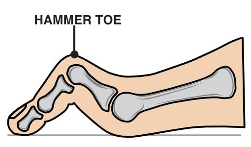
Hammer toe is a deformity that causes one or more of your toe joints to bend upward instead of lying flat.

Hammer toe affects the second, third, or fourth toe results from an imbalance in the muscles, tendons, and ligaments responsible for maintaining toe alignment. If left untreated, the affected toe may progress from being flexible to rigid, leading to increased pain and difficulty walking.

Anatomy of the Toes

The forefoot consists of five toes, each playing a crucial role in balance and movement.

Structurally, four of these toes have three joints: the metatarsophalangeal (MTP) joint at the base, the proximal interphalangeal (PIP) joint in the middle, and the distal interphalangeal (DIP) joint near the tip. However, the first toe, commonly known as the big toe, differs from the others as it has only two joints, the MTP and the interphalangeal (IP) joint.

In cases of hammer toe, the deformity primarily affects the PIP joint, causing it to bend abnormally while the other joints may remain straight or become misaligned over time.

What Is the Main Cause of Hammer Toes?

This condition often results from wearing ill-fitting shoes that force the toes into a bent position over extended periods.

As mentioned, hammer toe is the result of a soft tissue imbalance that puts pressure on the toe muscles, tendons and joints.

High heels or shoes that are too tight can force the foot down and push the toes against the shoe, increasing the pressure and the bend in the toe. If the toe is bent into one position of long enough, the muscles and joints begin to tighten and cannot be stretched out.

Additionally, certain medical conditions like rheumatoid arthritis or diabetes can increase the risk of developing hammer toes due to joint damage or nerve issues.

What are the Symptoms of Hammer Toe?

Hammer toe presents with several noticeable symptoms that can worsen over time.

Initially, you may experience mild discomfort and some hard skin developing on the toes, but as the condition progresses, symptoms can include:

Early recognition of these symptoms will help to ensure effective treatment and prevent longer-term complications.

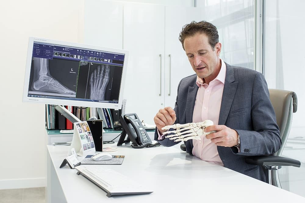
Diagnosis and Imaging Options

A clinical examination is the first step in diagnosing hammer toe. When you come into the clinic, I will assess your foot’s structure, flexibility, pain levels and mobility.

Imaging studies may be necessary for a more detailed evaluation:

It is very important to get an accurate and early diagnosis so that we can give you the most appropriate treatment for your specific symptoms and goals.

Can You Correct Hammer Toes?

Yes, hammer toes can be corrected. This can be done surgically or using non-surgical interventions depending on the severity.

Your symptoms and the flexibility of the affected toe play a significant role in determining the best and most effective treatment for you.

In the initial stages, mild hammer toe can be treated with non-surgical interventions when the toe is still flexible.

These may include:

These methods aim to alleviate symptoms and help prevent further progression of the deformity but are unlikely to correct the condition.

Unfortunately, if the toe has already become rigid then surgical intervention might be necessary to correct the deformity and to give you the best outcome in terms of pain relive, improved mobility and quality of life.

Do Hammer Toe Straighteners Really Work?

Hammer toe straighteners, such as splints or toe caps, can provide relief, especially in the condition's early stages. These devices help in:

It's important to note that while these devices can alleviate symptoms and provide temporary correction, they may not offer a permanent solution, particularly if the toe has become rigid.

How Do Podiatrists Treat Hammer Toes?

Podiatrists employ a range of treatments based on the severity of the hammer toe. Initial approaches often involve:

Your treatment will depend on an individual assessment and the progression of the deformity.

I work closely with expert podiatrists to provide a holistic and multidisciplinary approach to your treatment and care.

What Are the Surgical Options for Hammer Toes?

Surgical intervention may be necessary for patients with persistent or severe hammer toe.

Each surgical approach aims to relieve pain, improve toe function, and prevent any further complications.

The choice of procedure depends on whether the toe joint remains flexible or has become rigid.

It is then secured with an external K-wire or pin, which holds the bones in place while they fuse together.

Innovative Surgical Treatments for Hammer Toe

Advancements in medical technology have introduced innovative surgical treatments.

Instead of temporarily holding the operated toe in the corrected position with a pin, an internal implant can be used, which does not have to be removed. In certain situations, these implants are very beneficial.

Additionally, minimally invasive surgical techniques have been developed to correct hammer toes with smaller incisions, reducing recovery time and postoperative discomfort.

Is It Worth Getting Hammer Toe Surgery?

Deciding on whether to have surgery depends on several factors:

Surgical correction can offer long-lasting relief, improved toe function and better mobility, but I will always ensure that all the options are discussed, and we will make an informed decision together.

Surgical Patient

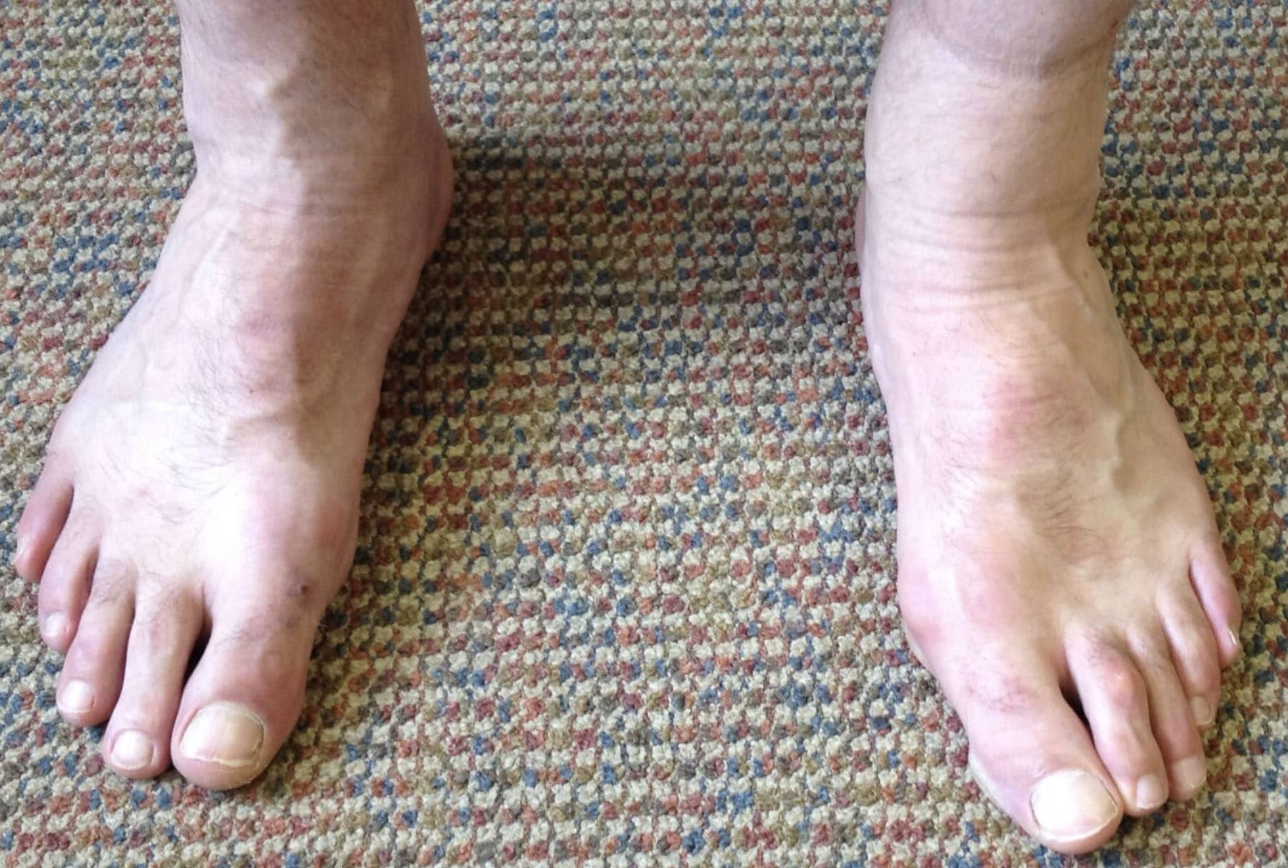
Post-operation hammer toe surgery, second toe on the right foot (left side of photo). The mild hammer toe on the other foot is being treated conservatively at present.

Is Hammer Toe Surgery Painful?

Hammer toe surgery is typically performed as a day case, so you are only in the hospital for a few hours and don’t usually stay overnight. The surgery is often performed under local anaesthetic rather than a general anaesthetic, although this will be discussed individually. You will not feel any pain during the procedure.

Postoperative discomfort varies among individuals, but we ensure that you receive the appropriate medication so you do not have pain.

You can weight bear after the operation and do not need crutches, but as hammertoe operations are often associated with after-forefoot operations, you might need to wear a stiff-soled post-op shoe to protect the operated foot.

It’s important that you follow the postoperative care instructions given to you, to ensure a smooth recovery.

How long does it take to recover from hammertoe surgery?

Recovery from hammer toe surgery varies based on the procedure performed. Initial skin healing from the surgery takes approximately 10-14 days.

Here are some guidelines to recovery timeframes:

Long-term outcomes are generally positive, and most patients experience significant pain relief and improved toe alignment and mobility.

Conclusion

Hammer toes are a common foot condition that can cause discomfort and affect daily activities. Early recognition and intervention are key to managing symptoms effectively and potentially avoiding surgery.

Whether through lifestyle modifications, non-surgical treatments, or surgical options, I will ensure that you receive a tailored approach to help you restore function and improve your quality of life.

Who Is the Best Doctor or Health Professional to See for Plantar Fasciitis?

Plantar fasciitis is one of the most common causes of heel pain, affecting people of all activity levels and ages. The condition stems from inflammation or microtears in the plantar fascia; a thick band of connective tissue running along the sole of your foot. While it often resolves with conservative treatment, knowing which healthcare professional to consult can significantly impact recovery time and outcomes.

As an Orthopaedic Foot and Ankle specialist, Plantar fasciitis is a condition that I see very regularly in my clinic, however, I believe that this condition is best managed by a multidisciplinary team of healthcare professionals, including GPs, podiatrists and Physiotherapists.

This blog explores the roles of different healthcare providers to help you decide who to see for plantar fasciitis.

Should I See a GP for Plantar Fasciitis?

General Practitioners (GPs) are often the first point of contact for any health issue, including plantar fasciitis.

A GP can:

A GP is a good starting point, particularly if you are unsure whether your heel pain is due to plantar fasciitis or another condition. However, they may lack the specialist expertise or tools to provide advanced treatment.

What Is the Best Practitioner for Plantar Fasciitis?

The "best" practitioner depends on your specific needs and the severity of your condition. Mild cases often resolve with basic self-care or guidance from a GP. However, more severe cases may require input from specialists such as podiatrists, physiotherapists, or orthopaedic consultants.

In general:

Who Is Best to Diagnose Plantar Fasciitis?

A proper diagnosis is key to effective treatment. Here’s how each professional approaches diagnosis:

GPs

A GP will have a low-level knowledge of this condition, they will likely make a diagnosis based on history and a physical exam, such as palpating the heel for tenderness and evaluating pain patterns.

Podiatrists

On top of what a GP is able to offer, a Podiatrist can also assess foot positions and biomechanics.

Physiotherapists

Physiotherapists will often look at the body as a whole and as well as focusing on the foot. They will evaluate movement patterns of the whole lower leg, gait (walking and running) and muscle imbalances; strengths and weaknesses that are likely contributing to the condition.

Orthopaedic Consultants

Orthopaedic Consultants perform advanced diagnostic imaging (e.g., MRI) if other conditions like heel spurs or stress fractures are suspected.

While GPs, Physios and podiatrists are well-equipped for initial diagnosis, more complex or unclear cases might benefit from imaging studies and specialist orthopaedic consultations and input.

When Should I See an Orthopaedic Consultant for Plantar Fasciitis?

Orthopaedic consultants specialise in surgical and non-surgical treatments for musculoskeletal conditions. You might consider seeing one if:

1.        Conservative Treatments Fail:

Persistent symptoms despite six months of treatments like stretching, orthotics, and shockwave therapy may warrant a specialist review.

2.        Advanced Imaging is Required:

If a precise diagnosis is unclear, orthopaedic consultants can order and interpret imaging like MRIs.

3.        Advanced Treatment is Needed:

An orthopaedic consultant is able to offer interventions such as extracorporeal shockwave therapy (ESWT) or steroid injections.

4.        Complex Foot Issues are Present:

If your plantar fasciitis is part of a broader foot problem, such as severe flat feet or tendon dysfunction, an orthopaedic consultant can provide comprehensive management.

5.        Surgery is Considered:

In rare cases, surgery to release the plantar fascia or remove a heel spur may be necessary.

The Role of Physiotherapists in Plantar Fasciitis Treatment

Physiotherapists play a pivotal role in the diagnosis and recovery process of plantar fasciitis pain, particularly when rehabilitation is required.

They will focus on the following:

·      Stretching and Strengthening Programme:

Targeted exercises for the calf muscles, Achilles tendon, and plantar fascia to alleviate pain and improve function.

·      Manual Therapy:

Techniques such as deep tissue massage and myofascial release can reduce tightness and improve flexibility.

·      Gait and Posture Analysis:

Physiotherapists can identify movement patterns that might be contributing to plantar fasciitis and recommend and facilitate appropriate adjustments.

·      Return-to-Activity Guidance:

They will help you gradually return to normal activities or sports without risking re-injury.

·      Offer Advanced Therapies:

Some Physios provide extracorporeal shockwave therapy (ESWT) or corticosteroid injections to address chronic cases.

Is It Worth Seeing a Podiatrist for Plantar Fasciitis?

Podiatrists are foot and ankle specialists, making them an excellent choice for plantar fasciitis. Their training allows them to:

If your symptoms persist despite initial treatments, or if you suspect that poor foot mechanics are making your condition worse, consulting a podiatrist can be highly beneficial.

What Order Should I See Professionals in for Plantar Fasciitis?

The sequence of care often depends on the severity and duration of your symptoms:

  1. GP (First Step):

For initial diagnosis and basic treatment advice.

If symptoms persist, these specialists can provide targeted treatments and advanced interventions.

For chronic or complex cases requiring advanced imaging or assessment and management.

By following this sequence, you ensure timely and appropriate care without jumping straight to invasive treatments.

Collaborative Care for Plantar Fasciitis

Effective management of plantar fasciitis often requires a multidisciplinary approach, as the condition can have multiple contributing factors such as biomechanical abnormalities, muscular imbalances, improper footwear, or overuse injuries. Collaboration between healthcare professionals ensures that all aspects of the condition are addressed, increasing the likelihood of a successful recovery.

The General Practitioner as the Coordinator

The GP typically serves as the first point of contact and acts as a coordinator for your care. They can refer you to the appropriate specialist based on the severity of your symptoms and your progress. For example, if conservative treatment such as rest, pain relief, and basic stretching does not improve your symptoms, the GP may involve a podiatrist or physiotherapist to focus on more specific interventions.

The Podiatrist and Physiotherapist Working Together

A podiatristand physiotherapistoften collaborate to provide comprehensive, holistic care. While the podiatrist focuses on correcting biomechanical issues, such as improper gait or lack of foot arch support, the physiotherapist addresses muscle imbalances and works on functional rehabilitation.

Orthopaedic Consultant for Advanced Cases

An orthopaedic consultant can provide more advanced imaging (such as ultrasound or MRI scan) to confirm the diagnosis and to rule out alternative conditions like painful heel spur, inflammation of the heel fat pad, nerve entrapment, stress fractures of the heel bone or even a rupture of the plantar fascia. 

The orthopaedic foot and ankle specialist works closely together with physiotherapists, podiatrists, and at times radiologists to design a treatment plan.

Conclusion

Plantar fasciitis can be a frustrating condition, but the right healthcare professional can make all the difference. Start with your GP for an initial evaluation and basic treatment. If symptoms persist, seek a podiatrist or physiotherapist for specialised care. Finally, consult an orthopaedic consultant for chronic or complex cases that may require advanced interventions. By taking a stepwise approach, you’ll maximise your chances of a full recovery and a fast return to pain-free activity.

Why Does My Heel Hurt? - Especially in the mornings

Solving the Mystery of Plantar Fasciitis – By Mr Martin Klinke

Plantar fasciitis is a prevalent and often debilitating condition that affects the bottom of the foot, primarily around the heel and arch of the foot.

As the leading cause of heel pain in the UK, plantar fasciitis impacts a wide demographic, from athletes to individuals with more sedentary lifestyles.

This article delves into the anatomy, causes, symptoms, risk factors, and treatment options for plantar fasciitis, giving some insight and helping those who are suffering from this condition to gain knowledge to be able to beat this common foot condition.

What is Plantar Fasciitis?

Plantar fasciitis is an overuse injury that occurs when the plantar fascia—a thick, fibrous band of tissue connecting the heel bone (calcaneus) to the toes—becomes irritated or inflamed. This tissue acts as a shock absorber, supporting the arch of the foot and playing a critical role in walking and running mechanics.

When excessive stress is placed on the plantar fascia, either due to a single event, or more usually, a low level of stress over a prolonged period of time, microtears can develop leading to inflammation, pain, and discomfort.

This condition typically develops gradually over a period of weeks or months, and is not always linked to a specific injury or trauma. Despite its prevalence, many sufferers delay seeking treatment, exacerbating the condition and prolonging recovery.

Anatomy of the Plantar Fascia

The plantar fascia is an important structural component of the foot, running from the heel bone to the base of the toes. It lies just beneath the skin on the sole of the foot and serves several vital functions:

Due to its constant involvement in movement and weight-bearing activities, the plantar fascia is highly susceptible to injury.

Causes of Plantar Fasciitis

Plantar fasciitis is a multifactorial condition with no single definitive cause. However, several contributing factors can increase your susceptibility to developing it.

1. Overuse or Overloading

2. Footwear Choices

3. Weight Gain

4. Biomechanical Factors

5. Age and Tissue Degeneration

6. Medical Conditions

Symptoms of Plantar Fasciitis

The hallmark symptom of plantar fasciitis is a pain on the bottom of the heel, often described as sharp, stabbing, or throbbing. This pain typically worsens when walking or running, especially after a period of rest.

Other symptoms may include:

Diagnosing Plantar Fasciitis

A thorough clinical examination is essential for diagnosing plantar fasciitis.

Key diagnostic steps include:

Treatment Options for Plantar Fasciitis

Treatment aims to reduce pain, promote healing, and prevent recurrence. Approaches can be categorised into self-care, conservative therapies, advanced interventions, and surgical options.

1. Self-Care Measures

For the calf stretch make sure that your heel is down on the floor. Hold for 30-45 seconds and repeat 3 times.

You can use a tennis or golf ball to gently massage and stretch the plantar fascia, along the length of your foot arch.

These can be done 2-3 times a day

2. Pain Management

You should seek professional advice before starting any medication

3. Physiotherapy and Podiatry

A physiotherapist can design a tailored exercise program to improve flexibility and strengthen supporting muscles. Common techniques include:

4. Shockwave Therapy

Extracorporeal shockwave therapy (ESWT) is a non-invasive treatment that delivers powerful sound waves to the plantar fascia. Benefits include:

Studies show positive outcomes after 3-5 sessions of ESWT.

5. Ultrasound-Guided Injections

For persistent pain, injection therapy may be considered:

6. Surgical Interventions

Surgery is very rare and reserved for severe, refractory cases. Options include:

Preventing Plantar Fasciitis

Preventive strategies are crucial to reduce the likelihood of developing plantar fasciitis or experiencing a recurrence:

When to Seek Professional Help

While self-care measures are often relatively effective, certain situations warrant medical attention:

Plantar fasciitis can significantly impact your quality of life, but with timely intervention and proper management, most individuals experience substantial improvement.

If you’re struggling with the relentless heel pain and discomfort of plantar fasciitis, I’m here to help you take the first steps toward relief.

As a dual-trained foot and ankle surgeon with a degree in sports medicine, I bring a unique perspective to your care. I don’t just look at your foot in isolation—I consider how your entire body moves and functions, particularly if you’re active or trying to get back to the sports and activities you love.

My goal is to help you recover fully, regain your mobility, and leave heel pain behind. I also have a team of Physiotherapists and Podiatrists who I work closely with, and can therefore ensure a holistic approach to your treatment.

Together, we’ll create a tailored approach to get you back on your feet and thriving!

Case Study: Morton’s Neuroma in a Recreational Runner – Surgical Intervention and Recovery

Presentation

A 32-year-old avid runner presented in my clinic with a longstanding Morton’s neuroma, diagnosis (diagnosed by her GP and Physiotherapist) that had significantly impacted her ability to train and participate in endurance events. Her symptoms had initially been managed with conservative treatment, including changing her footwear, physiotherapy and reducing her running, but these interventions ultimately failed to provide long-term relief. Her frustration with persistent pain led her to seek further consultation.

History and Conservative Treatments

This lady initially managed her neuroma symptoms with wider-fit footwear, avoiding doing up her laces too tightly on the running shoes, and reducing her training, all of which seemed to give mild reduction in her symptoms, but did not resolve them.

She went on to have some Physiotherapy, which focused on reducing tightness in her calf muscles, working on her balance and proprioception, and looking at her walking and running technique. She also tried some off the shelf insoles which only seemed to exacerbate her symptoms.

Minimally Invasive Treatment

In our first consultation we explored all her options to manage her symptoms.

Together we decided to initially try a corticosteroid injection, which is a very safe and common treatment option for Moreton’s neuroma. This initially brought significant pain relief, and the patient was able to get back to her normal life and running training. However, as is often the case, the symptoms returned within a few months, leaving her frustrated and still unable to enjoy running or even walking without discomfort.

Decision for Surgical Intervention

After exhausting conservative treatments and given her clinical history, and the fact that her life and mental wellbeing was being so negatively affected, we had an open discussion about surgery.

While the idea of an operation can be daunting, this lady felt ready for a solution that could offer a long-term return to her active lifestyle. We decided that a neurectomy (a procedure to remove the thickened nerve), would give her the best chance at lasting relief.

The patient opted for surgery which was covered through her private health insurance.

Surgical Approach

The procedure was done as a day case surgery, was performed under general anaesthetic. The Morton’s neuroma was excised along with a significant portion of the nerve, extending to the mid-arch region. This extended excision approach helps prevent the formation of a “stump neuroma,” which could reintroduce symptoms if the nerve were only partially removed. Post-operatively, she was given a surgical sandal and crutches and instructed on pain management, including avoiding opioids if possible.

Postoperative Recovery and Rehabilitation

The surgery went smoothly, and within days the patient noticed a marked difference. The usual pain was gone, replaced by mild, manageable post-operative soreness.

During the early recovery phase, I emphasised the importance of elevating the foot to reduce swelling. The patient’s partner provided extensive support, handling daily tasks to allow for an easier recovery time.

By two weeks, she returned for a follow-up; while healing was progressing well, she still had some residual nerve sensitivity and shooting pain when attempting to walk in clinic, a common response as the nerve heals. At this stage I referred her to Physiotherapy for post operative rehabilitation.

Her recovery was then managed through progressive physiotherapy, focusing initially on reducing nerve sensitivity and restoring functional gait. Physiotherapy interventions included gait training with one crutch, controlled load-bearing activities, and gradual strengthening exercises specific to her biomechanics.

Key to her recovery was a gradual reintroduction to impact-based activities, such as walking and low-impact gym exercises, leading to more advanced exercises like stationary cycling and rowing.

At approximately eight weeks post-operation, she began a carefully monitored return-to-run program. This consisted of brief run-walk intervals on grass to minimise impact. Her progress was monitored weekly, with steady improvements observed in foot strength, gait alignment, and endurance.

Outcome and Long-term Prognosis – Back to an Active Lifestyle

A few months post-surgery, the patient was back on her feet in every sense of the word. She returned to running and hiking and was finally free from the nagging nerve pain that had disrupted her daily life.

She completed a 25K trail race 10 months after the surgery which included challenging terrain, with no neuroma-related pain during or after the event.

The patient remains highly satisfied with her surgical outcome, experiencing minimal discomfort and demonstrating an encouraging return to long-distance running. She anticipates training for future marathons and remains mindful of incorporating gradual progression to avoid flare-ups.

Conclusion

This case underscores the potential of surgery as an excellent option for treating Morton’s neuroma when conservative measures are no longer effective.

When managed appropriately, surgery can offer a durable long-term solution; and with careful postoperative management and guidance from a sports-focused physiotherapist, patients can successfully return to high-impact activities.

I would encourage any patient considering surgery to discuss expectations and timelines thoroughly, as recovery varies depending on each individual.

If you’re struggling with this painful condition and conservative treatments aren’t providing relief, it is worth exploring surgical options. Life is too short to be held back by pain, especially for those who thrive on an active lifestyle.